


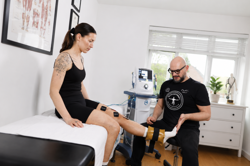


Piotr Kolodziej | Specialist Physiotherapist | PK Physiotherapy, Northampton
Introduction
Finding out that you have a torn meniscus can be worrying, particularly if your work involves kneeling or you want to get back to football.
A common concern is whether the knee can recover without surgery. In many cases, a structured rehabilitation programme is an appropriate starting point. However, some injuries need an early orthopaedic opinion, especially when a displaced fragment blocks movement or there is a tear that may benefit from repair. [3, 4]
The decision depends on the type of damage, the condition of the rest of the knee and what you need to return to. It also helps to distinguish two different outcomes: the meniscus physically healing and the knee becoming comfortable and functional again.
In this article, I explain the main injury patterns, what physiotherapy can achieve, when surgery becomes appropriate and what current research and UK guidance actually tell us.
What Is the Meniscus?
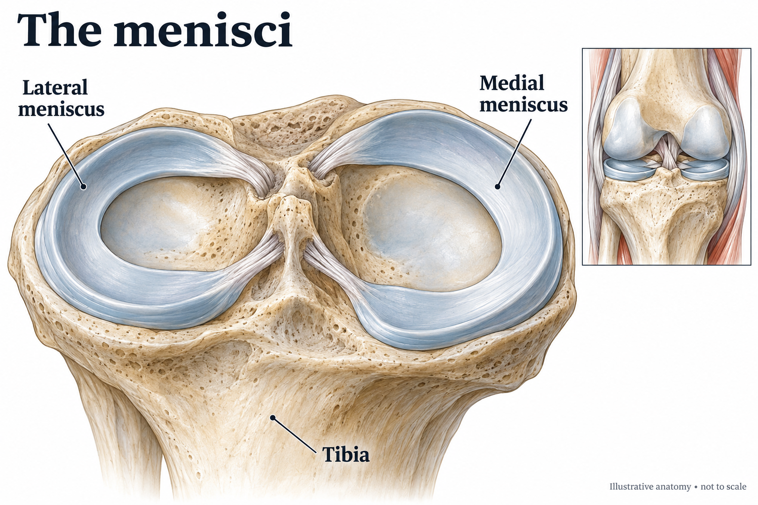
Each knee has two menisci: the medial meniscus on the inner side and the lateral meniscus on the outer side.
These curved structures are made from fibrocartilage and sit between the thigh bone and shin bone. They spread load across the joint and contribute to stability. They are different from the smooth articular cartilage covering the ends of the bones. [1]
Preserving useful meniscal tissue matters. Removing part of it changes how the knee distributes load, which is one reason modern surgical guidance emphasises preservation whenever appropriate. [3]
Traumatic and Degenerative Meniscal Injuries
Traumatic Tears
A traumatic tear follows an identifiable injury, often involving a loaded knee twisting during a turn, tackle or awkward landing. A footballer may have the foot planted while the body rotates.
The meniscus can be injured alone or alongside a ligament, particularly the ACL. Assessment therefore needs to consider the whole knee. Associated injuries can change both the treatment decision and the rehabilitation plan. [16]
Degenerative Meniscal Changes
Degenerative lesions develop as tissue quality changes over time. Symptoms may begin gradually or after a relatively minor movement, such as standing from a squat. They frequently coexist with osteoarthritis.
A degenerative tear on MRI does not automatically establish the cause of pain. The scan needs to be interpreted alongside symptoms, examination and the condition of the surrounding joint. [4, 5]
Different Types of Meniscal Tear
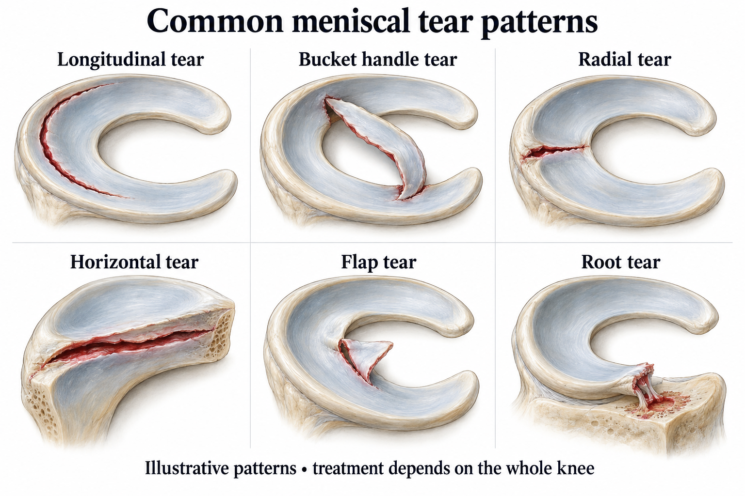
Longitudinal Tears
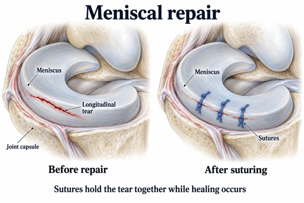
These run along the direction of the meniscal fibres. Some peripheral tears are suitable for repair, depending on stability and tissue quality.
Bucket Handle Tears
A longer fragment can displace towards the centre of the joint. This may prevent the knee from straightening and requires prompt assessment.
Radial Tears
These cut across the fibres involved in transmitting load. Large or complete radial tears can substantially compromise meniscal function and may need specialist consideration for repair. [16]
Horizontal Tears
These split the tissue into upper and lower layers and are commonly associated with degeneration.
Flap and Complex Tears
A flap tear creates a partly detached piece of tissue. A complex tear combines more than one pattern. Their significance depends on symptoms, stability and the overall knee. [1, 2]
Meniscal Root Tears
The roots anchor the meniscus to the shin bone. A root tear can seriously affect its ability to distribute load, even when the damaged area appears relatively small.
These injuries deserve specialist assessment. Repair may be appropriate in selected patients, but cartilage condition, alignment and the wider clinical picture affect the decision. A suspected root tear should not automatically be treated as an ordinary degenerative lesion. [2, 16]
What Does a Meniscal Injury Feel Like?
Symptoms may include pain around the joint line, swelling, stiffness, discomfort with twisting or squatting, and difficulty fully bending or straightening the knee. Some people notice catching or a feeling that the knee may give way. [14]
Symptoms alone cannot identify the exact tear pattern. Being able to walk does not exclude an injury, and clicking alone does not establish that surgery is needed.
Catching and a Truly Locked Knee
Brief catching is different from a knee that remains physically blocked and cannot move normally. A persistently locked knee needs urgent assessment. Do not repeatedly force it straight or try to exercise through the obstruction. [4, 5]
Seek urgent GP advice or NHS 111 help if you cannot bear weight after an injury, have severe knee pain, cannot bend or straighten the leg, or develop a hot swollen knee with fever or feeling unwell. A deformed knee or altered sensation after injury needs A&E assessment. [14]
How Is a Meniscal Injury Assessed?
Assessment begins with the history: how the problem started, whether there was a clear injury, when swelling appeared and which movements are difficult.
The examination may assess movement, joint line tenderness, ligament stability and functional tasks. Clinical tests such as McMurray or Thessaly can contribute to the assessment, but no single test should be treated as a complete diagnosis. [3]
It is also important to consider other explanations for knee pain. For somebody who kneels at work, discomfort may involve a bursa, tendon or the joint itself rather than an isolated meniscal problem.
You can read more about assessment on the PK Physiotherapy Knee Pain page.
Do I Need an MRI?
MRI is useful when it is likely to change management. EBI guidance highlights significant acute injuries with mechanical symptoms, locked knees and persistent symptoms despite initial care as situations where referral and imaging may be appropriate.
For many people with gradual symptoms and no concerning features, treatment can begin without an immediate scan. Imaging is particularly important when a repairable injury is suspected. [5]
Can a Torn Meniscus Heal on Its Own?
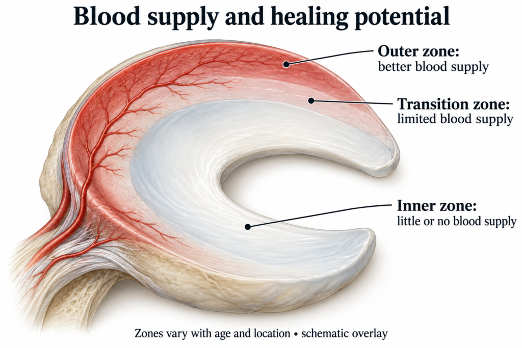
Some tears can heal without surgery, especially small, stable tears towards the better supplied outer edge. The inner part has much less blood supply and a poorer capacity for healing. [1]
Clinicians sometimes describe a red zone, a transitional red and white zone, and a white zone. These refer to differences in blood supply. They help explain healing potential but do not provide a guaranteed outcome for an individual tear. [2]
Healing and Feeling Better Are Different
The knee can become substantially more comfortable and functional even if the tear remains visible on MRI. Better strength, movement and load tolerance can make daily life easier without proving that the tissue has rejoined.
This distinction matters when interpreting research. Trials showing good results from rehabilitation primarily measure symptoms and function. They do not establish that exercises physically close every tear. [6, 7]
There is no responsible single percentage for the chance of spontaneous healing across all injuries. A stable peripheral tear, a displaced bucket handle tear and a degenerative lesion represent different clinical situations.
The Science Behind Physiotherapy for Meniscal Injuries
Physiotherapy aims to restore the knee’s capacity for useful activity while respecting the injury. Rest alone does not rebuild the strength required for stairs, lifting equipment or slowing down during a football match.
Strength and Physical Capacity
Progressive resistance exercise develops the muscles supporting the lower limb. In the trial by Kise and colleagues, supervised exercise produced greater improvement in thigh muscle strength at three months than partial meniscectomy. At two years, the main patient reported outcome showed no clinically relevant difference between groups. [9]
The practical implication is that a rehabilitation programme needs a meaningful training component. A series of appointments without appropriate exercise progression is not equivalent to the programmes tested in research.
Movement Control and Graded Exposure
Balance and movement practice help prepare the leg for tasks that require control. Loading can progress from daily activities towards the demands of work or sport, with symptoms and performance guiding the next step. Education helps the patient understand the plan and recognise when reassessment is needed. These elements are supported by the international meniscus rehabilitation consensus. [11]
Physiotherapy does not replace a detached root or remove a fragment that is mechanically obstructing the joint. Those problems require a different decision pathway. [2, 4]
What Does the Research Show?
The ESCAPE Trial
The ESCAPE trial included 321 people aged 45 to 70 with degenerative meniscal tears. At five years, exercise based physiotherapy remained noninferior to arthroscopic partial meniscectomy for knee function, within the trial’s predefined margin.
This supports rehabilitation as the preferred initial option for many similar patients. However, the trial excluded locked knees and injuries requiring acute surgery. Its findings should not be applied indiscriminately to every traumatic tear. [6]
The FIDELITY Trial
FIDELITY compared partial meniscectomy with placebo surgery in people with a degenerative medial meniscal tear. At five years, meniscectomy offered no relevant benefit in symptoms or function and was associated with a slightly greater risk of radiographic osteoarthritis progression.
This challenges routine trimming of degenerative tears. It does not show that repairing an appropriate acute injury is ineffective: repair is a different procedure. [8]
The STARR Trial
STARR studied 100 people aged 18 to 45 with an isolated traumatic tear. Early partial meniscectomy was not superior at two years to physiotherapy with the option of later surgery.
In the physiotherapy group, 41% ultimately underwent delayed surgery during follow up. The remaining 59% did not. The study excluded locked knees and tears considered suitable for suturing on MRI, which limits how broadly its conclusions can be used. [7]
The DREAM Trial
An exploratory analysis of DREAM, published in 2024, found no apparent advantage of early surgery over exercise and education for pain, function and quality of life after 12 months in either the traumatic or nontraumatic subgroup.
Because this was an exploratory subgroup analysis, it should support a balanced discussion rather than a universal rule against surgery. [10]
What Evidence Based Practice Means
Evidence based practice combines relevant research, clinical assessment and the patient’s goals. It also recognises where evidence is weaker.
Confidence is strongest for rehabilitation as an initial approach in many degenerative cases. Decisions about particular acute tear patterns require more individual judgement. Trials of partial meniscectomy must not be presented as trials of meniscal repair. [6, 7, 8]
What Does Nonoperative Treatment Involve?
Managing the Early Symptoms
After an acute injury, temporarily reduce activities that clearly aggravate symptoms. This may include loaded twisting, prolonged kneeling and deep squatting. Gentle movement can be reintroduced as tolerated after the initial rest period rather than keeping the knee completely still.
Cold packs wrapped in a towel may help symptoms. Ask a pharmacist or doctor about suitable pain relief, particularly if you take other medicines or have relevant health conditions. [14]
Building a Rehabilitation Programme
The programme should connect exercises to practical goals. Depending on assessment, examples might include:
• gentle knee movement and quadriceps activation
• controlled sit to stand practice
• step ups and progressive squat variations
• resistance exercises for the thigh, calf and hip
• balance and single leg control
• later practice of work or sporting tasks
These are examples, not a prescription for every tear. Exercise range, resistance and frequency need to be individualised. A postoperative repair may require restrictions that do not apply to someone receiving nonoperative care. [9, 13]
I would review how the knee responds during activity and afterwards. More swelling, a worsening limp or reduced movement the following day would prompt an adjustment. This is a practical way of monitoring load, not proof that the tissue is safe under every exercise.
When Should the Plan Be Reviewed?
Progress should be assessed regularly. If symptoms persist despite an appropriate programme, reconsider the diagnosis, loading and need for further investigation.
EBI uses approximately three months of initial nonoperative care as a review point for persistent symptoms in relevant patients. It is not a mandatory waiting period for a locked knee or a potentially repairable acute injury. [4, 5]
What Does NICE Guidance Say?
NICE NG226 concerns osteoarthritis in people over 16. It is relevant when degenerative meniscal changes form part of an arthritic knee, but it is not a guideline for every acute sports tear.
Recommendations 1.3.1 to 1.3.4 support tailored therapeutic exercise, consideration of supervised sessions and education. Weight management is relevant where appropriate. Manual therapy should only be considered alongside exercise for hip or knee osteoarthritis.
Recommendation 1.7.1 advises against arthroscopic lavage or debridement for osteoarthritis. Recommendation 1.3.9 advises against treatments including TENS, interferential therapy, ultrasound and laser for osteoarthritis because evidence of benefit is insufficient.
These recommendations should not be misrepresented as a ban on surgery for a separate repairable injury. EBI and specialist meniscal guidance address additional questions about referral and treatment selection. [12]
Read the official NICE NG226 recommendations.
When Should a Knee Surgeon Be Consulted?
An early opinion matters when there is a displaced tear restricting movement or an acute injury with repair potential. Persistent disabling symptoms despite appropriate care may also justify referral.
The aim of consultation is to clarify the options. It does not commit the patient to surgery. Useful questions include whether repair is possible, what benefit is expected and how the postoperative restrictions would affect work or family life. [3, 4]
Meniscal Repair
Repair brings the torn edges together using sutures or fixation devices, allowing an opportunity for healing while preserving tissue. For a root repair, the surgeon may anchor the attachment using sutures passed through a small tunnel in the shin bone. [2]
Partial Meniscectomy
This removes an unstable fragment that cannot appropriately be preserved. It does not reconnect the tear. When surgery is indicated, retaining as much functioning meniscal tissue as possible is an important principle. A quicker initial recovery after trimming is not, by itself, a reason to remove repairable tissue. [3]
What Happens During Arthroscopy?
Arthroscopy is a surgical technique using small incisions. A camera allows the surgeon to view the inside of the joint on a screen, while instruments are introduced through other openings.
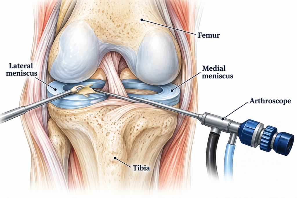
The anaesthetic is discussed beforehand. Knee arthroscopy may be performed under general or spinal anaesthesia. The surgeon examines the joint and carries out the agreed procedure where indicated. The incisions are then closed and dressed. Many patients go home the same day. [15]
What Is Arthroscopy?
Arthroscopy, also called diagnostic arthroscopy, means looking directly inside the joint to investigate a problem. The term does not mean that a meniscus will automatically be repaired or removed.
Any possible treatment during the same operation should be discussed as part of informed consent. Ask what question the procedure is intended to answer and why examination and imaging have not provided enough information. [15]
Simply looking inside the knee does not guarantee relief. Routine arthroscopic washing or clearing out a degenerative knee should not be presented as a proven solution to osteoarthritis. [8, 12]
Arthroscopy still carries risks, including infection, blood clots, stiffness, persistent symptoms and anaesthetic complications. A repair may fail to heal or be injured again. [2, 15]
Rehabilitation Before and After Surgery
Before Surgery
Where surgery is planned and there is no reason for urgent intervention, preparation can focus on comfortable movement, appropriate strength work and understanding the postoperative restrictions. A blocked knee should not be forced through exercises or have urgent assessment delayed for a routine rehabilitation course.
Practical planning matters too: using crutches, managing stairs, arranging transport and modifying work may all need discussion.
After Partial Meniscectomy
Weight bearing and movement can usually progress more quickly than after repair, according to symptoms and the surgical team’s advice. Rehabilitation still needs to address swelling, movement, strength and functional control. [13]
After Meniscal Repair
Repair requires protection while healing takes place. Crutches, a brace or limits on bending and loading may be needed. The exact restrictions depend on the tear and operation, so the physiotherapist needs the operative information.
Root, radial and more complex repairs may require a more protective or prolonged programme. A generic arthroscopy exercise sheet is not enough to determine suitable progression. [13]
Returning to Sport
The international consensus gives broad return to sport ranges of 4 to 12 weeks after partial meniscectomy and 6 to 9 months after repair. These are guidance ranges, not guaranteed deadlines.
Readiness also involves symptoms, movement, strength, functional performance and confidence. A footballer needs to tolerate running, braking, turning and training before match play. Associated ACL injury may substantially change the pathway. [11]
For that situation, read ACL Injuries Explained.
How I Approach Meniscal Injuries at PK Physiotherapy
At PK Physiotherapy in Northampton, assessment and treatment are provided personally. I want to understand what happened, what is limiting you now and what a useful recovery would look like in your daily life.
For a handyman, that may mean getting down to floor level, carrying tools and completing a working day. For a Sunday League footballer, it includes tolerating unpredictable turns and the fatigue of a match.
I use the assessment to guide an individualised exercise programme and review whether the plan is producing worthwhile progress. The programme needs to be manageable outside appointments, with clear advice about activity and what should trigger reassessment.
Where Do Hands On Treatment and Physical Modalities Fit?
The availability of a treatment does not make it necessary for every injury. I would not describe massage, shockwave, laser, ultrasound or electrotherapy as proven ways to reconnect a torn meniscus.
Any additional treatment needs a specific clinical purpose and an honest discussion of its evidence. It should not displace active rehabilitation or delay a necessary orthopaedic opinion. Where osteoarthritis is present, the NICE recommendations above are particularly relevant. [12]
If surgery has been performed, post surgical rehabilitation must reflect the actual procedure and the surgeon’s restrictions. “Arthroscopy” alone does not tell us whether tissue was inspected, trimmed or repaired.
Meniscal Injury Case Studies
The following examples are based on patients I have treated at PK Physiotherapy. Individual details have been kept general to protect patient confidentiality. Treatment was adapted to the findings at assessment and to each patient’s functional goals.
Case Study 1: A Handyman Working on His Knees
I treated a 52 year old handyman who developed gradually increasing pain along the inner side of his knee. His work involved frequent kneeling, getting up and down from the floor, carrying tools and working for prolonged periods in restricted positions.
There had been no major twisting injury and the knee was not locked. His main difficulties were pain when rising from kneeling, stiffness after work and discomfort with deeper knee bending.
My assessment focused not only on the possibility of meniscal irritation, but also on knee movement, joint line symptoms, muscle strength, patellofemoral loading and the surrounding soft tissues.
The main part of his treatment was progressive rehabilitation. We worked on quadriceps and hip strength, controlled sit to stand exercises, step exercises, functional squatting and gradually improving his ability to move safely between standing and floor level.
I also used manual therapy to improve comfortable knee movement and reduce stiffness. Soft tissue massage was used where surrounding muscles were tight and contributing to discomfort. IFC was used as an additional method of short term pain management during the early stages of rehabilitation. Kinesiology tape was also used temporarily when it helped the patient feel more comfortable during activity.
Importantly, these additional treatments were not presented as methods of physically repairing a torn meniscus. Their role was to help manage symptoms and allow the patient to participate more effectively in active rehabilitation.
We also discussed changes at work. These included reducing prolonged kneeling where possible, changing position more frequently, avoiding loaded twisting from a deep kneeling position and breaking demanding tasks into shorter periods.
As his strength and tolerance improved, the exercises became increasingly specific to his work. This included lifting, carrying, controlled squatting and repeated transitions between standing and floor level.
Over the course of rehabilitation his pain reduced, knee function improved and he became progressively more comfortable completing his normal working duties.
The important point was not whether an MRI abnormality had disappeared. The practical outcome was that his knee became stronger, more tolerant of load and better able to cope with the demands of his job.
Case Study 2: A Sunday League Footballer
I treated a Sunday League footballer who developed knee pain following a twisting movement while changing direction during a match.
The knee became swollen afterwards and he reported pain with turning, deeper bending and some movements involving rotation. He was still able to walk and there was no persistent mechanical locking of the knee.
The initial assessment included knee movement, swelling, joint line symptoms, ligament stability, strength and functional control. Because there was no true locked knee or other indication for urgent surgical management, we started a structured rehabilitation programme.
During the early stage I concentrated on reducing irritation, restoring comfortable movement and maintaining muscle activation. IFC was used for short term symptom management where appropriate, and kinesiology tape was used temporarily to provide additional support and improve comfort during activity.
Manual therapy was used where restriction in knee movement was present, together with soft tissue treatment for surrounding muscles.
The central part of treatment was exercise.
We gradually progressed from quadriceps activation, controlled knee flexion and basic strengthening to single leg exercises, balance work and more demanding lower limb strengthening.
Once the knee tolerated these exercises without increased swelling or significant next day symptoms, rehabilitation became more football specific.
We introduced running progression, acceleration and deceleration work, single leg loading and eventually controlled changes of direction.
The final stages included turning, braking, football specific movement patterns and preparation for returning to team training.
I did not use pain relief treatments as evidence that the meniscus had healed. They were used only as supportive treatments while strength, movement and load tolerance were rebuilt through rehabilitation.
The player returned gradually rather than going directly from pain free walking to a full match. Training tolerance, knee response, strength and confidence were considered before unrestricted football.
This case demonstrates an important point in meniscal rehabilitation. A football player does not automatically need surgery simply because the injury occurred during a twisting movement. The decision depends on the type of tear, whether the knee is mechanically blocked, associated injuries and how the knee responds to appropriate rehabilitation.
My Clinical Perspective
The most useful treatment plan gives the patient a clear explanation and a practical route forward.
I want someone to leave understanding why we are choosing rehabilitation, why further investigation might be necessary, and what progress would look like. If the knee is improving, the programme should help build on that improvement. If it is not, we should reconsider the plan.
There is a place for both physiotherapy and surgery in meniscal care. The decision should be specific enough to explain why a particular approach suits this person and this knee.
Q&A
Does Every Meniscal Tear Need Surgery?
No. Many people can begin with rehabilitation. Displaced tears, persistent locking and injuries that may benefit from repair require a different discussion. [3, 4]
Can I Walk With a Torn Meniscus?
Often, provided walking is tolerable and there are no restrictions. Inability to bear weight after injury needs urgent assessment. Follow the prescribed loading limits after repair. [13, 14]
Does Clicking Mean the Tear Is Getting Worse?
Clicking alone cannot establish that. Increasing pain, recurrent swelling or a new loss of movement is a reason for reassessment. Persistent locking needs prompt attention. [4, 5]
Will Physiotherapy Make the Tear Disappear on MRI?
That is not a reliable promise. Rehabilitation can improve function without demonstrating structural closure of the tear. [6, 7]
Should I Avoid Squats Permanently?
There is no universal lifetime ban. Range and resistance can be adjusted to the individual. After repair, progression must also respect tissue protection and the surgical protocol. [11, 13]
Will a Steroid Injection Heal the Meniscus?
No. An injection may be considered for symptom management in selected circumstances, but it does not reconnect the tear. [2]
Will the Meniscus Grow Back After Trimming?
You should not expect the removed portion to regrow into a normal meniscus. Preservation is therefore important when an operation is needed. [3]
How Long Should I Try Physiotherapy?
There is no single period that suits everyone. Progress and tear characteristics matter. Persistent symptoms warrant review, while a locked knee or potentially repairable acute injury should not be put through an automatic waiting period. [4, 5]
Can I Return to Kneeling at Work?
This depends on symptoms, capacity and the type of treatment. Following repair, kneeling may remain restricted while the tissue heals. A graded return is usually more useful than testing a full working day at once. [13]
Does Surgery Guarantee a Pain Free Knee?
No. The condition of the whole joint matters. In degenerative tears, trimming may offer no additional benefit over rehabilitation, whereas selected acute injuries have different indications. [6, 8]
Book Your Physiotherapy Assessment
If knee pain is affecting work, kneeling, stairs or sport, an assessment can help clarify the problem and the next step.
At PK Physiotherapy, we can discuss your symptoms, examine the knee and develop an appropriate plan, including whether an orthopaedic opinion is needed.
You can also read about Knee Pain, Sports Injuries, Post Surgical Rehabilitation and Specific Personalised Exercise Programmes.
This article provides general information and does not replace an individual assessment. A locked knee or the other urgent symptoms described above needs prompt medical attention.
Library
Medical evidence and further reading. References and linked destinations checked on 25 September 2026. Numbers in the article refer to the sources below. Detailed rehabilitation recommendations include expert consensus as well as trial evidence, and the certainty is not equal for every tear pattern.
[1] AAOS OrthoInfo. Meniscus Tears. Patient information on anatomy, symptoms and healing potential. Read the AAOS information.
[2] AAOS OrthoInfo. Meniscus Repair. Indications, surgical techniques and recovery. Read about meniscal repair.
[3] AAOS. Management of Acute Isolated Meniscal Pathology, 2024. This guideline does not cover chronic or degenerative lesions, root tears or tears with associated ACL injury. Guideline scope and access. AAOS summary of recommendations.
[4] Academy of Medical Royal Colleges. EBI guidance on arthroscopic surgery for meniscal tears. Reviewed September 2024. Read the EBI recommendations.
[5] Academy of Medical Royal Colleges. EBI guidance on knee MRI for suspected meniscal tears. Reviewed September 2024. Read the MRI guidance.
[6] Noorduyn JCA and colleagues. ESCAPE randomised clinical trial, five year follow up. JAMA Network Open. 2022;5:e2220394. DOI 10.1001/jamanetworkopen.2022.20394. Read the publication record.
[7] van der Graaff SJA and colleagues. Arthroscopic partial meniscectomy versus physical therapy for traumatic meniscal tears in a young study population. British Journal of Sports Medicine. 2022;56:870 to 876. STARR randomised trial. Read the publication and DOI.
[8] Sihvonen R and colleagues. FIDELITY placebo surgery controlled trial, five year follow up. British Journal of Sports Medicine. 2020;54:1332 to 1339. Read the publication and DOI.
[9] Kise NJ and colleagues. Exercise therapy versus arthroscopic partial meniscectomy for degenerative meniscal tear in middle aged patients. BMJ. 2016;354:i3740. DOI 10.1136/bmj.i3740. Read the BMJ article.
[10] Damsted C and colleagues. Early surgery versus exercise therapy and patient education for traumatic and nontraumatic meniscal tears in young adults. Exploratory analysis from the DREAM trial. Journal of Orthopaedic and Sports Physical Therapy. 2024. DOI 10.2519/jospt.2024.12245. Read the publication record.
[11] Prill R and colleagues. The Formal EU US Meniscus Rehabilitation 2024 Consensus. Part II, prevention, nonoperative treatment and return to sport. Published 2025. Read the consensus.
[12] NICE. NG226, Osteoarthritis in over 16s: diagnosis and management. 2022. Relevant recommendations include 1.3.1 to 1.3.4, 1.3.7, 1.3.9 and 1.7.1. Read the official NICE guidance.
[13] The Formal EU US Meniscus Rehabilitation 2024 Consensus. Part I, rehabilitation after meniscectomy, repair and reconstruction. Published 2025. Read the postoperative rehabilitation consensus.
[14] NHS. Meniscus tear. Symptoms, self care and when to seek urgent help. Read the NHS information.
[15] NHS. Arthroscopy. What the procedure involves, recovery and potential complications. Read about arthroscopy.
[16] Kopf S and colleagues. Management of traumatic meniscus tears: the 2019 ESSKA meniscus consensus. Published 2020. Read the consensus.
Useful PK Physiotherapy Links
Initial Physiotherapy Assessment
Specific Personalised Exercise Programmes